

A group from the civil engineering department at the University of Waterloo (Ontario, Canada) have demonstrated a novel, imaging-driven technique that provides high accuracy quantification of cyanobacteria. The technique, which appeared in Nature Scientific Reports in June, is intended to demonstrate that high accuracy, imaging based, rapid water quality analysis can be achieved with conventional equipment available in typical water quality laboratories.
The method seemingly “identifes and enumerates cyanobacterial cells at a level equivalent to or better than that achieved using standard manual microscopic enumeration techniques, but in less time, and requiring signifcantly fewer resources.”
When compared with indirect measurement methods, the proposed method is said to provide better accuracy at both low and high cell concentrations. It extends the detection range for cell enumeration while maintaining accuracy and increasing enumeration speed.
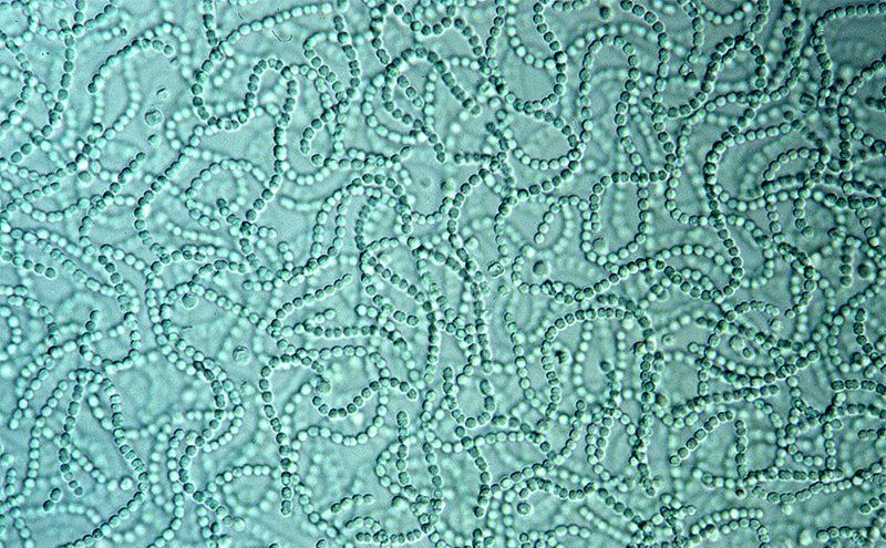
The developed method not only accurately estimates cell concentrations but it also reliably distinguishes between cells of Anabaena fos-aquae, Microcystis aeruginosa, and Ankistrodesmus in mixed cultures by taking advantage of additional contrast between the target cell and complex background gained under fluorescent light.
So this image-driven approach seems to offer promise as a robust and cost-effective tool for identifying and enumerating microscopic cells based on their unique morphological features.
As the paper explains, cost effective, fast, and reliable cyanobacterial cell identification and enumeration methods are much in demand, for water quality monitoring programs. The paper reviews a number of direct and indirect analytical methods that are available. For example, direct microscopic enumeration using a hemacytometer is the most commonly identifcation method. Indirect quantification methods have also been developed but these tend to require expensive equipment. For example, flow cytometry, PCR-fuorescent fragment detection, and in-situ fuorescence.
Imaging-based enumeration methods present a number of advantages for rapid and low-cost water quality monitoring of cyanobacteria, including in relation to their cost effectiveness and speed, their specificity (because they can detect and differentiate target cells based on size, shape, and colour), and the fact that they can be readily integrated into current monitoring methods. These methods are also automated, customisable and offer flexibility when it comes to developing quality control and assurance protocols.
Nonetheless, such methods still present technical challenges that are a barrier to the realisation of automated cell enumeration techniques that can be used routinely for water quality monitoring. The paper explains how the proposed method overcomes some of these problems.